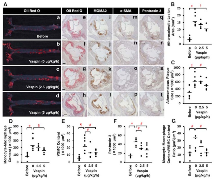
Figure 6.
Effects of vaspin on atherosclerotic lesion development in Apoe−/− mice. Seven mice were sacrificed before infusion (17 weeks old), and 7, 5, and 6 mice were sacrificed after a 4-week continuous subcutaneous infusion of 3 dosing rates of vaspin (0, 2.5, 5 μg/kg/h), respectively. (A) Atherosclerotic lesions were stained with Oil Red O on the aortic surface (a–d). Cross-sections of the aortic sinus were stained with Oil Red O (e–h), MOMA2 for monocytes/macrophages (i–l), α-SMA for VSMCs (m–p), or pentraxin 3 for vascular inflammation (q–t). Hematoxylin was used for nuclear staining. Bar = 200 μm. (B–G) Comparisons of these positive area and the ratio of monocyte-macrophage content/VSMC content within atheromatous plaques were performed among 4 groups. Bars indicate the mean values in the graphs. * p < 0.0001, † p < 0.005, ‡ p < 0.001, # p < 0.05, § p < 0.01, ¶ p < 0.0005.

