Figure 1.
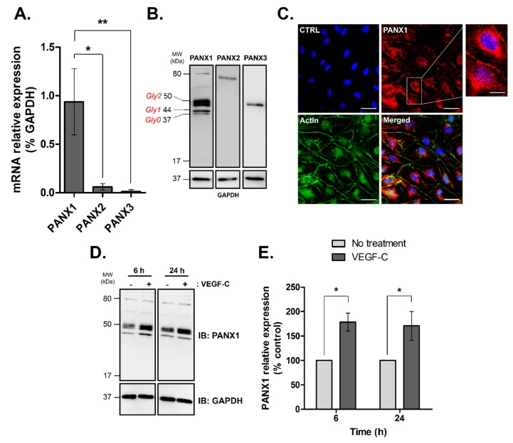
Pannexin isoforms expression in human dermal lymphatic endothelial cells (HDLECs). (A) PANXs mRNA expression in isolated HDLECs quantified by RT-PCR and normalized by GAPDH. The data represent mean ± SD from three independent experiments; (B) Western blot analysis of total protein extracts (20 μg/lane) from four independent HDLEC cultures demonstrating PANXs expression in HDLECs. Unglycosylated (Gly0) and glycosylated isoforms (Gly1 and Gly2) of PANX1 are indicated; (C) PANX1 immunofluorescence in HDLECs (red), F-actin was FITC-phalloidin stained (green) and nuclei were DAPI-stained (blue). CTRL: control immunofluorescence after omission of the primary antibody, Scale bar: 50 μm; Enlarged image marked by the white box shows higher magnification of PANX1 staining, scale bar 7 µm; (D) Representative Western blot analysis and (E) densitometric quantification of PANX1 expression normalized to GAPDH following 100 ng/mL VEGF-C treatment for the indicated times in HDLECs. Values are expressed as mean ± SD from three independent experiments. * p < 0.05 and ** p < 0.01.

