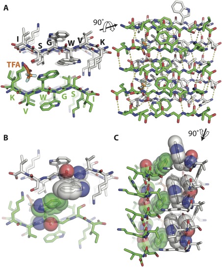
Figure 3.

Segment 30‐KVWGSI‐35 of SOD1 forms a steric zipper assembly. (A) The 1.45 Å resolution structure of segment KVWGSI shows two β‐sheets composed of antiparallel β‐strands forming a class 7 steric zipper via face to back stacking. Shown here in views parallel (left) and perpendicular (right) to the fibril axis. Trifluoroacetic acid (TFA) is colored orange. (B) The two sheets do not pack tightly due to the bulky tryptophan side chain in the inner interface. (C) In most steric zippers, side chains are perpendicular to the protofilament axis but here, the W32 side chain protrudes along the fiber axis and aligns with the G33 of the β strand above it as shown in sphere representation. This results in staggering of the sheets relative to each other.
