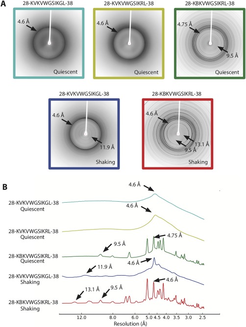
Figure 5.

Comparison of diffraction patterns of the different variant segments. (A) Top: 28–38 and G37R mutant segments show diffuse diffraction rings at 4.6 Å. The G37R segment forming out‐of‐register pairs of sheets reveals sharp diffraction rings at 4.75 and 9.5 Å. Bottom: The segment 28–38 under shaking conditions reveals diffraction rings consistent with cross‐β structure, indicative of pairs of sheets. (B) Radial profiles of the diffraction patterns with peaks at or around 4 and 11 Å labeled.
