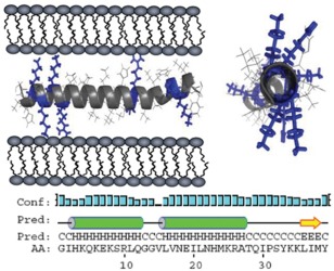
Figure 3.

Illustration of SEVI‐induced membrane fusion. Two views of a model α‐helical structure of PAP248–286 showing the distribution of positively charged residues, and the predicted secondary structure of PAP248–286 within full‐length PAP. The letter codes “C, H, and E” denote a residue in random coil, helical, or β‐sheet conformation, respectively. Figure adopted from a previously published article.28
