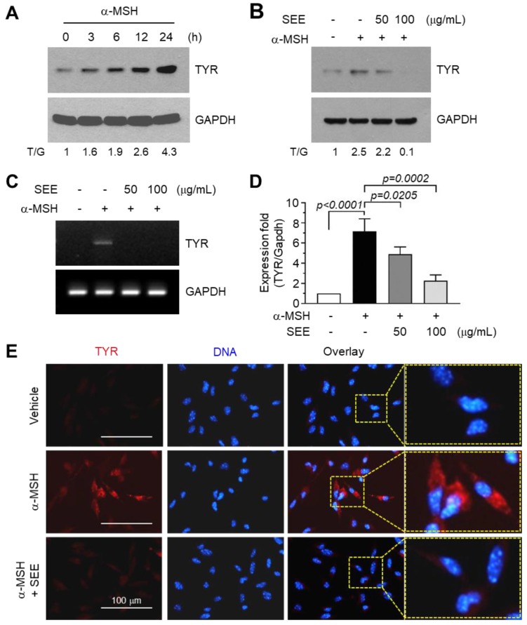
Figure 1.
Effect of sorghum ethanolic extract (SEE) on the suppression of alpha-melanocyte-stimulating hormone (α-MSH)-induced tyrosinase (TYR) expression. (A) B16F10 cells were treated with 100 nM α-MSH for various times (0–24 h) and cell lysates were subjected to immunoblotting using antibodies against TYR. The glyceraldehyde 3-phosphate dehydrogenase (GAPDH) level was examined as an internal control. T/G, tyrosinase/GAPDH (B) B16F10 cells were treated with either vehicle (DMSO) or SEE (50 and 100 μg/mL) for 30 min, followed by stimulation with 100 nM α-MSH. After 12 h, cell lysates were prepared, and immunoblotting was performed using antibodies against TYR. The GAPDH level was examined as an internal control. The intensity of the bands was quantified using ImageJ and the relative TYR intensity was normalized to that of GAPDH and visualized in the blot. T/G, TYR/GAPDH. (C,D) B16F10 cells were treated with either vehicle (DMSO) or SEE (50 and 100 μg/mL) for 30 min, followed by stimulation with 100 nM α-MSH. After 6 h, total RNA was isolated and TYR mRNA was measured by RT-PCR (C) and quantitative real-time PCR (D). The GAPDH mRNA level was examined as an internal control. (E) B16F10 cells were treated with vehicle (DMSO) or SEE (50 μg/mL) in the absence or presence of 100 nM α-MSH. After 12 h, the cells were fixed and incubated with antibodies against TYR for 2 h, followed by incubation with Alexa Fluor 555-conjugated (red signal) secondary antibodies for 30 min. Nuclear DNA was stained with 1 μg/mL Hoechst 33258 for 10 min (blue signal). The dotted box indicates the region of higher magnification in images.

