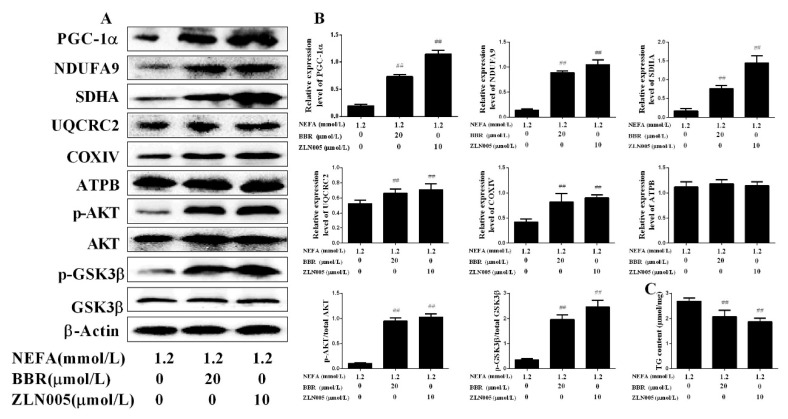
Figure 8.
Activation of PGC-1α increased the beneficial effects of BBR on mitochondrial respiratory chain function and insulin signaling damage induced by NEFA. Hepatocytes were assigned to 3 groups as follows: A 1.2 mmol/L NEFA group, a 1.2 mmol/L NEFA + 20 μmol/L BBR treatment group, and a 1.2 mmol/L NEFA + 10 μmol/L ZLN005 treatment group. (A,B) Western blot analysis and quantification of key molecules of the insulin signaling pathway, PGC-1α, and five representative subunits of OXPHOS complexes, and β-Actin served as an internal control; (C) TG content in bovine hepatocytes. Quantified data are mean ± SD; ## p < 0.01 versus NEFA group.

