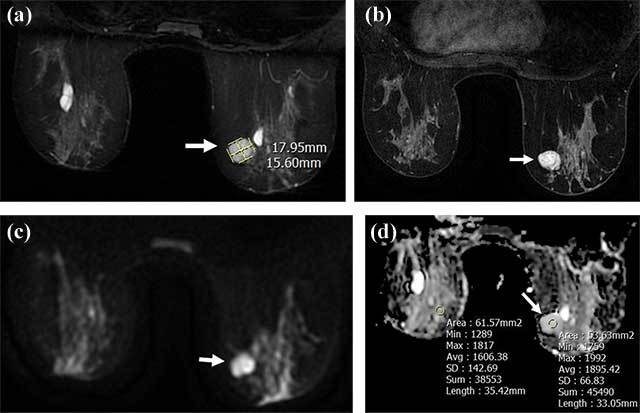
Figure 1.

A well-circumscribed 18 × 15.6 mm mass lesion in the right breast upper-inner quadrant hyperintense (arrow) in T2-weighted images (a), homogeneously enhancing (arrow) in T1-weighted post-contrast images (b) without diffusion restriction in DWI (c, d); ADC and rADC are 1.895 and 1.179, respectively (d). Tru-cut biopsy results revealed fibroadenoma.
