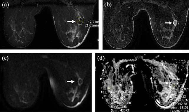
Figure 2.

A 18 × 22 mm irregular mass lesion in the right breast lower-outer quadrant, hypointense (arrow) in T2-weighted images (a) with rim enhancement (arrow) in contrast images (b), showing diffusion restriction (arrow) in DWI; ADC and rADC values are 0.955 and 0.544, respectively (c, d). Tru-cut biopsy results diagnosed invasive ductal carcinoma.
