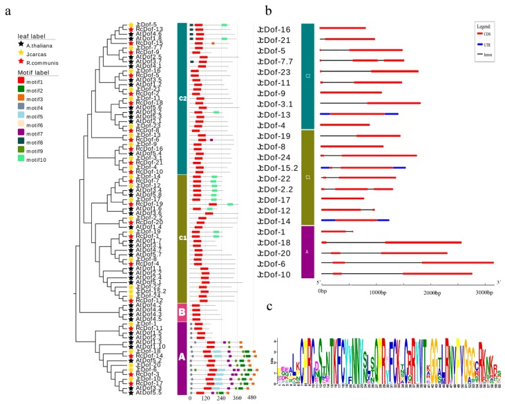
Figure 3.
JcDof gene structures and conserved motifs in JcDof proteins. (a) The distribution of 10 conserved motifs in Dof proteins; (b) Gene structures of JcDof genes. CDS, UTR and introns were depicted by filled red boxes, blue boxes, and single black lines; and (c) Motif-1, corresponding to theCX2CX21CX2C single zinc-finger structure. The detailed motif’s sequences are shown in Figure S1 and Table S4.

