
Figure 3.
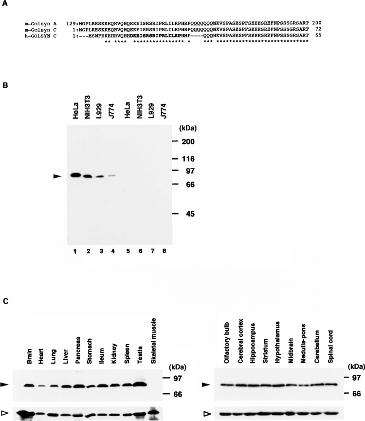
Expression of m-Golsyn protein in mouse cell lines and tissues. (A) Parts of the peptide sequences of m-Golsyn and h-GOLSYN are indicated. The peptide sequence in bold type was used for the preparation of the anti-h-GOLSYN antibody. (B) Extracts (20 μg/lane) of HeLa, NIH3T3, L929, and J774 cells were separated by SDS-PAGE on a 7.5% slab gel and immunoblotted with anti-GOLSYN antibody in the absence (lanes 1–4) or presence (lanes 5–8) of an excess amount of the peptide used for the preparation of anti-GOLSYN antibody. (C) Lysates (50 μg/lane) prepared from various mouse tissues were separated on a 7.5% SDS-PAGE and analyzed by immunoblotting with anti-GOLSYN antibody as described in Materials and Methods. Left panel: various tissues including brain; right panel: various regions of the central nervous system. Solid and open arrowheads indicate the positions of m-Golsyn protein and β-tubulin, respectively.