
Figure 4.
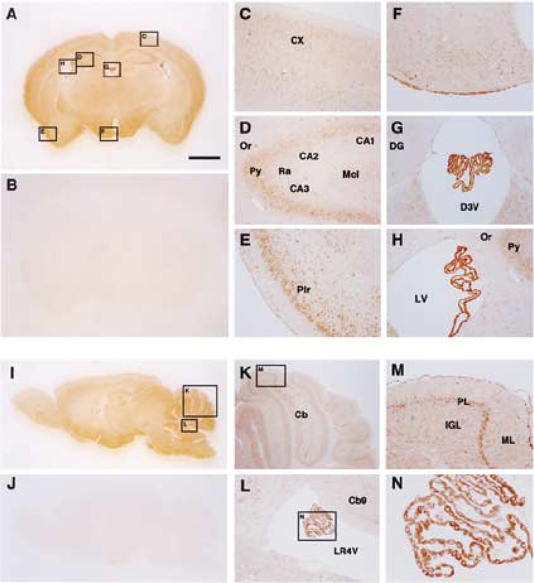
Immunohistochemical analysis of m-Golsyn protein in mouse brain. Coronal (A–H) and sagittal (I–N) sections of the mouse brain were stained with anti-GOLSYN antibody in the presence (B and J) or absence (A to N except B and J) of an excess amount of the peptide used for the preparation of anti-GOLSYN antibody. Regions indicated by boxes C to H in (A) are magnified and shown in (C) to (H), respectively. Boxes K and L in (I) are magnified and shown in (K) and (L), respectively. CX, cerebral cortex; Or, oriens layer of the hippocampus; Py, pyramidal cell layer of the hippocampus; CA1, CA2, and CA3, CA1, CA2, and CA3 areas of the hippocampus; Ra, stratum radiatum of the hippocampus; Mol, molecular cell layer of the hippocampus; Pir, pirform cortex; DG, dendate gyrus; D3V, third ventricle; LV, lateral ventricle; Cb, cerebellum; LR4V, fourth ventricle; PL, Purkinje cell layer of the cerebellum; ML, molecular layer of cerebellum; IGL, inner granular layer of cerebellum. Scale bar: 1 mm.