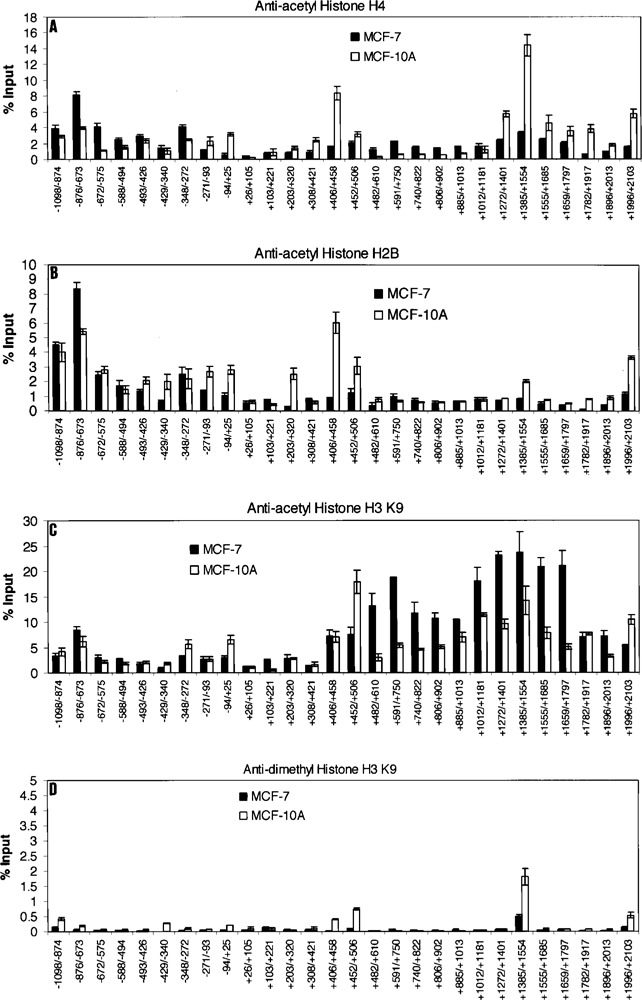
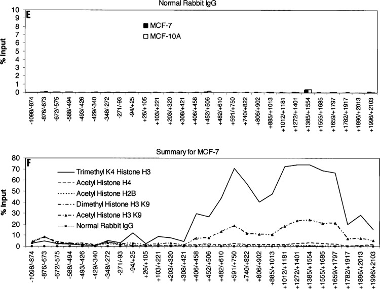
Figure 3.


Histone modifications within the Keratin 8 gene locus in MCF-7 and MCF-10A cells. ChIP assays were performed with anti-acetyl histone H4 (A), anti-acetyl histone H2B (B), anti-acetyl histone H3 K9 (C), or anti-dimethyl histone H3 K9 (D). Normal Rabbit IgG served as the negative control (E). The amount of precipitated DNA was quantified relative to input as described in Materials and Methods. All data are presented as the means ± SD of two independent experiments. The initiation site of TATA box is designated at +1. (F) Summary and comparison of the histone modifications examined on the Keratin 8 gene in MCF-7 cells.
