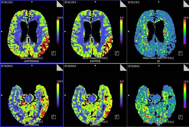
Figure 2.

Top (a) and bottom (b) slices of brain perfusion CT shows extensive perfusion alterations in the entire left parietotemporal region, consisting of a normal to slightly diminished MTT (right column), and an increased CBF (left column) and CBV (middle column).
