Figure 3.

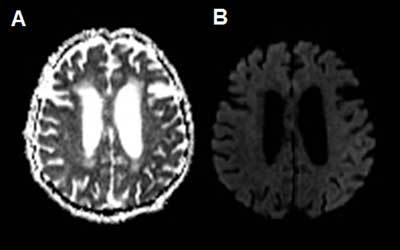
ADC (a) and b1000 DWI (b) show subtle diffusion restriction in the cortex of the left parietotemporal lobe, corresponding with the hyperperfused region observed on perfusion-CT.

ADC (a) and b1000 DWI (b) show subtle diffusion restriction in the cortex of the left parietotemporal lobe, corresponding with the hyperperfused region observed on perfusion-CT.