
Figure 1.
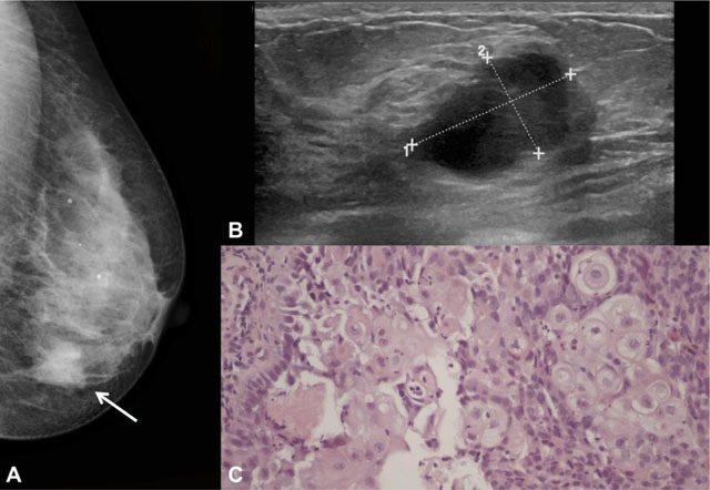
Example of squamous cell subtype in a 66-year-old woman. A – Left craniocaudal mammogram showing an irregular dense mass in the inner quadrants of the breast (arrow), with non-circumscribed margins. B – At ultrasound, it corresponded to a partially circumscribed breast mass with complex solid and cystic echo structure and posterior acoustic enhancement. C – Photomicrograph showing squamous cell differentiation (H&E, 200x).