
Figure 2.
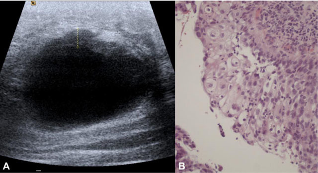
Large left breast mass in a 36-year-old woman with squamous cell subtype. A – Sonogram demonstrating a large cystic lesion with irregular anterior wall thickening. B – Photomicrograph showing squamous cell differentiation (H&E, 200x).
Official websites use .gov
A
.gov website belongs to an official
government organization in the United States.
Secure .gov websites use HTTPS
A lock (
) or https:// means you've safely
connected to the .gov website. Share sensitive
information only on official, secure websites.
Large left breast mass in a 36-year-old woman with squamous cell subtype. A – Sonogram demonstrating a large cystic lesion with irregular anterior wall thickening. B – Photomicrograph showing squamous cell differentiation (H&E, 200x).