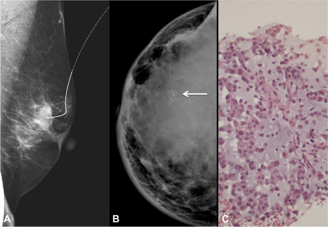
Figure 3.

A – Left mediolateral oblique mammogram of a 68-year-old woman with spindle cell subtype. Pre-operative needle for localization in a partially circumscribed oval dense mass. B – Right craniocaudal mammogram of a 30-year-old woman with matrix producing subtype, showing a global asymetry with coarse calcifications (arrow). There is also associated skin thickening (especially in the inner quadrants) and nipple retraction. C – Photomicrograph of the same lesion in B demonstrating the presence of chondromyxoid matrix (H&E, 200x).
