
Figure 1.
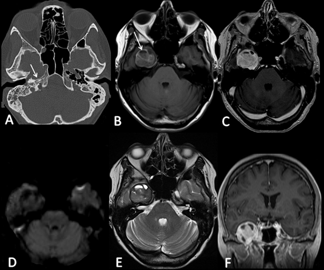
A) CT demonstrates a well-defined petrous lacunar image (arrow). B) T1-weighted image shows the hyperintense round lesion centered on the right Meckel cavum, relying on the intrapetrous carotid artery, and generating a mass effect on the temporal lobe (arrow). D) DWI is negative. E) T2-weighted image shows the presence of cystic areas. C) and F) Axial and coronal T1-weighted images show a slightly heterogeneous enhancement after contrast administration.