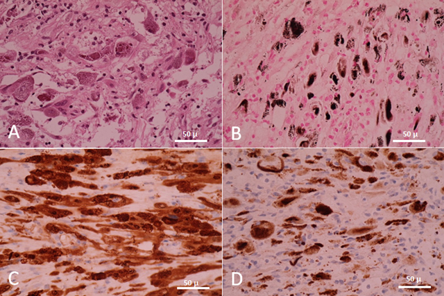
Figure 2.

Microscopic pictures of tumor cells (×40) reveal their dual Schwann and melanocytic phenotype. A) Hematoxylin-eosin staining shows plump pigmented tumorous Schwann cells. No psammomatous bodies are observed. B) Fontana staining shows the presence of melanin in tumorous Schwann cells. C) S-100 protein and D) HMB-45 confirm the diagnosis.
