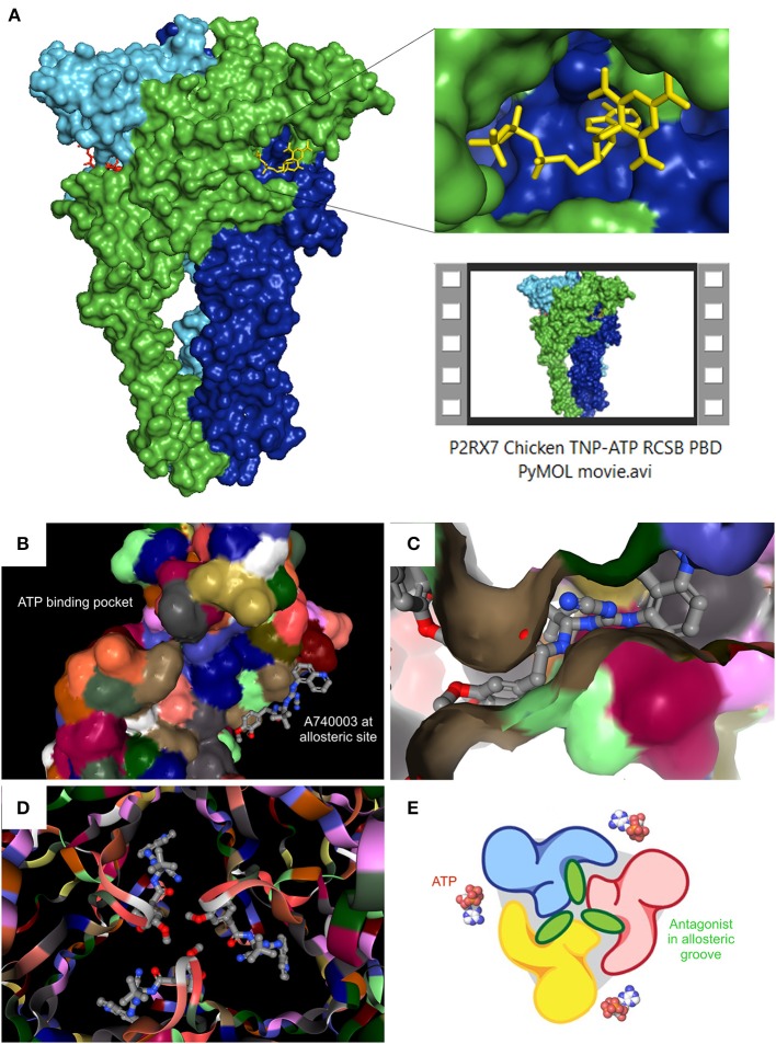
Figure 1.
Recent P2RX7 structural developments: the ATP binding pocket and the allosteric groove. (A) PyMOL (pymol.org) generated surface plot of P2RX7 trimer using RCSB PDB data file (rcsb.org) for chicken variant bound to the competitive antagonist TNP-ATP (structure 5XW6, 3.1A). ROI shows the ATP binding pocket (RHS, upper), PyMOL Video file gives structural overview of the trimer with detailed orientations of the ATP binding pocket and the central ion channel (see Supplementary Material Video S1). RCSB PDB generated images show location of the ATP binding pocket in relation to the newly discovered allosteric site on the monomer (B) and at the subunit interface of the trimer, forming an allosteric groove—here shown occupied by A740003 antagonist (C). When imaged together from intracellular projection, the three allosteric grooves appear as a Shuriken, whose rotation is impeded by the allosteric antagonist A740003 (D). (E) Cartoon representation of the relationship between the agonist binding site and the allosteric antagonist-binding groove. Adapted from Karasawa and Kawate (2016), with permission.

