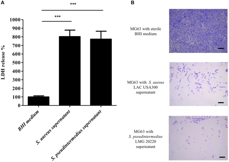
Figure 1.
Effect of S. pseudintermedius supernantant on NPPc (MG63). MG63 cells were incubated with staphylococcal supernatants for 4 h at 37°C. (A) Quantification of LDH release, reflecting NPPc lysis by staphylococcal supernatant. All of the results are expressed as the percentages of the values obtained for the control (BHI medium alone, 100%). Bars represent means ± standard deviations derived from 3 experiments performed in triplicate. The difference in the LDH concentration in staphylococcal supernatants condition compared to BHI medium alone was evaluated using a one-tailed Mann-Whitney test with a α risk of 0.05 (***p < 0.001). (B) MG63 lysis after 4 h in contact with staphylococcal supernatant. The cells were stained with Giemsa, and observed for morphological changes by light microscopy at a magnification of ×20. Bars, 500 μm. LDH, Lactate dehydrogenase; NPPc, Non-professional phagocytic cells.

