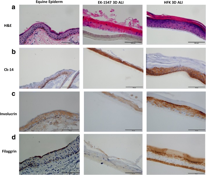
Fig. 7.
The air-liquid interface of primary equine epithelial cells recapitulate the in vivo skin epithelium. To assess the ability of equine keratinocytes to form a stratified epidermis in ALI (middle panel) morphology was compared to normal equine skin (left panel) by hematoxylin and eosin (H&E) (a) and stratification marker CK14 (b) involucrin (c) and filaggrin (d). HFK in ALI was used as technical control. All images (×40 magnification, scale bar = 100 μm) are representative of three experimental repeats

