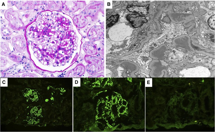
Figure 2.
Proliferative GN with monoclonal Ig deposits, IgM variant. This figure describe an IgM variant of the proliferative GN with monoclonal Ig deposits which was initially described only with monoclonal IgG deposits. (A) Light microscopy exhibits a mesangial proliferative GN pattern of injury (periodic acid–Schiff stain). (B) On electron microscopy, granular mesangial and subendothelial electron dense deposits (without substructure) are seen. On immunofluorescence, there was bright granular glomerular mesangial positivity for (C) IgM and (D) κ-light chain with negative glomerular staining for (E) λ-light chain. Glomeruli were negative for IgG and IgA (not shown). Magnification, ×400 in A, D, and E; ×4800 in B; ×200 in C.

