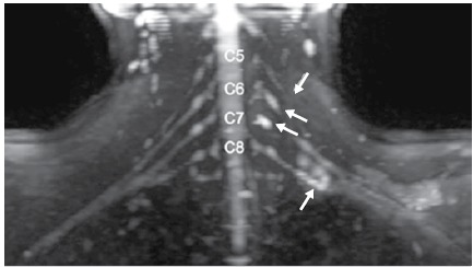
Figure 6. Magnetic resonance imaging, patient 3. Diffusion-weighted neurography - reconstruction in the coronal plane: Thickening and increased signal in the C5 root and region of the division of the lower trunk. Formation of pseudo-meningocele in the emergence of the C6 and C7 roots, suggestive of avulsion (pre-ganglionic lesion).

