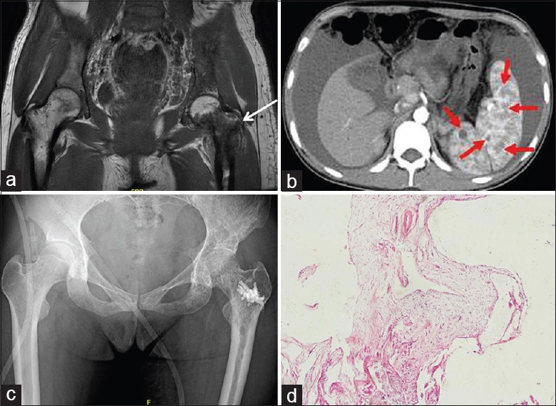
Figure 1.

Gorham–Stout syndrome of the left femur treated with cement augmentation in a 27-year-old female. (a) Preoperative coronal T1-weighted fat-saturated magnetic resonance imaging scan revealed the bony destruction indicated by arrows. (b) Transverse plane of the computed tomography scan revealed cystic lesions in the spleen. (c) Postoperative posteroanterior radiograph of the hip showed cement augmentation was satisfactory. (d) Histopathology revealed dilated lymphatic channels in hyperplastic fibrous connective tissue with lymphocytic infiltrate among the trabeculae (H and E staining, original magnification ×40).
