
Figure 3.
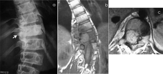
Osteoblastoma of T11. Radiograph (a) shows scoliosis and focal sclerosis of T11 (arrow). Coronal T1-WI (b) and axial T1-WI after gadolinium contrast administration (c) shows a lesion of the neural arch of T11, intralesional calcification and adjacent bone marrow edema of the vertebral bodies of T10 and T11. Note the compression of spinal cord (arrowhead).