
Figure 12.
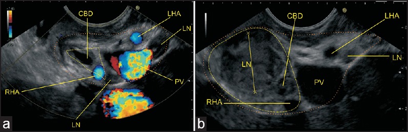
The HDL contains the structures of the portal triad. In Figures a and b, the yellow triangle demarcates the lower and upper part of hepatocystic triangle within the HDL. The orange line demarcates the area of HDL. A lymph node (LN) belonging to the pericholedochal group is seen. The pathological LN belongs to the cystic group of node