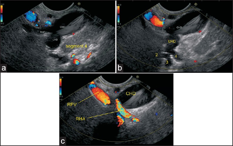
Figure 5.

(a) Imaging from the duodenal bulb can show the CBD/ CHD in a long axis close to the probe. The union of RHD (average length 0.9 cm) and left hepatic duct (average length 1.7 cm) to form the CHD can be seen (b) This union is seen close to the right end of the porta in front of the right branch of the PV. The RHA crosses behind the CHD to come and lie in the hepatocystic triangle (c) Sometimes, it is possible to see the segmental ducts to segments II, III, and IV from the hilum of the liver
