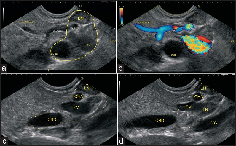
Figure 7.

(a) A transpancreatic imaging of the CBD and PV is performed in this case. The hepatoduodenal ligament is seen from the stomach between the upper border of the pancreas and the visceral surface of the liver (b) Two LNs belonging to the superior pancreaticoduodenal group are also seen (c) The PHA is seen entering the hepatoduodenal ligament, along with the PV and supra duodenal part of the bile duct (d) The intrapancreatic part of the bile duct is seen, which is not included in the HDL. The foramen of Winslow node is seen
