
Figure 8.
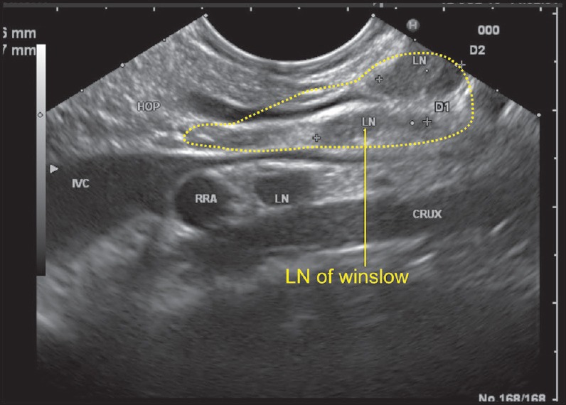
As the scope is positioned near the fundus of the stomach, it is able to see the inferior vena cava (IVC) and PV in a long axis. The IVC passes posterior to the hepatoduodenal ligament while the PV exits from the upper border of the pancreas and enters the lower part of the hepatoduodenal ligament. In this position, the bile duct can be also seen entering the ligament from the pancreas and the foramen of Winslow can be imagined as a place between the IVC and the PV just above the upper border of the pancreas. A lymph node of the foramen of Winslow is seen posterior to the PV and anterior to the IVC