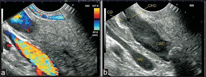
Figure 9.

(a) When the imaging is performed from the duodenal bulb, the PV can be followed up from the upper border of the pancreas into the hepatoduodenal ligament (b) The CBD divides into the CHD and cystic duct. The cystic duct joins the right aspect of the common hepatic duct. This union generally lies above the pancreas in the hepatoduodenal ligament
