Figure 2.

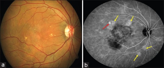
Color fundus photograph (a) of a patient with polypoidal choroidal vasculopathy. Midphase indocyanine green angiography (b) showing the presence of polyp (Red arrow) and multifocal areas of hyperfluorescence (Yellow arrows)

Color fundus photograph (a) of a patient with polypoidal choroidal vasculopathy. Midphase indocyanine green angiography (b) showing the presence of polyp (Red arrow) and multifocal areas of hyperfluorescence (Yellow arrows)