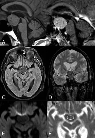
Figure 2.

Sagittal T1-weighted MR images before (A) and after (B) gadolinium administration demonstrating a somewhat inhomogeneous aspect with moderate to intense enhancement of the tumor. Axial flair (C) and coronal T2-weighted (D) images showing the occupation of the opto-chiasmatic cistern and the low signal intensity to gray matter of the lesion. Diffusion-weighted image (E) demonstrating a slight hypointensity with ADC value estimated at 0.70 × 10⁻³ mm2/s (F)(circle).
