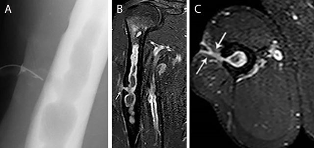
Figure 13.

Chronic osteomyelitis of the right humerus with fistula formation. Plain radiograph (sinography) (a), coronal (b) and axial (c) Fat–Sat T1–WI after gadolinium contrast administration. The sinography (a) shows an intramedullary well defined lytic lesion with scalloping of the cortex. Note the presence of a catheter in the sinus. There is enhancement of the wall of the intra-osseous abscess and the wall of the fistula (white arrow) (b, c). Image courtesy Dr. H. Declercq, Dendermonde.
