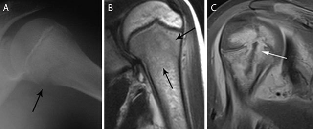
Figure 3.

Acute osteomyelitis in 2 different patients. Plain radiograph (a) and coronal T2–WI (b) of acute osteomyelitis in the left proximal humerus. Another example of a child with osteomyelitis in the right proximal humerus (c) on coronal T1–WI with FS after gadolinium contrast administration. Standard radiography (a) shows a subtle osteolytic lesion at the metaphysis and loss of cortical delineation of the medial humerus (black arrow). The surrounding bone marrow edema (black arrows) (b) is restricted to the metaphysis in most cases of childhood osteomyelitis. The relativity of the barrier of the growth plate on MRI is illustrated in (c) Although a rim-enhancing intra-osseous abscess (white arrow) corresponding with the main site of infection is located at the metaphysis, there is also focal enhancement of the growth plate and epiphysis, in keeping with metaphyseal crossing.
