
Figure 1.
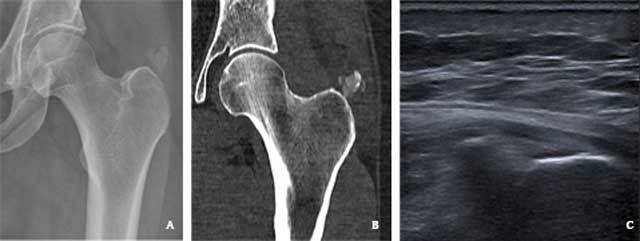
A 46-year-old woman with calcific tendinitis of the left gluteus medius tendon. (A) Plain radiography reveals a large calcification superior to the greater trochanter (B) CT with soft tissue window confirms this calcification (arrow), and locates it in the gluteus medius tendon. (C) Ultrasound image confirms a hyperechogenic calcification in the gluteus medius tendon.