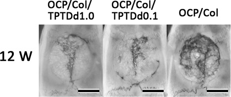
Figure 3.

Radiographic examination. In the OCP/Col/TPTDd1.0 and OCP/Col/TPTDd0.1 groups, most of the defect is covered by a radiopaque figure, which is denser, more granulous, and has relatively uniform radiopacity. In OCP/Col, most of the defect is occupied by radiopacity, which consists of the fusion of small radiopaque masses. Bars: 4 mm.
