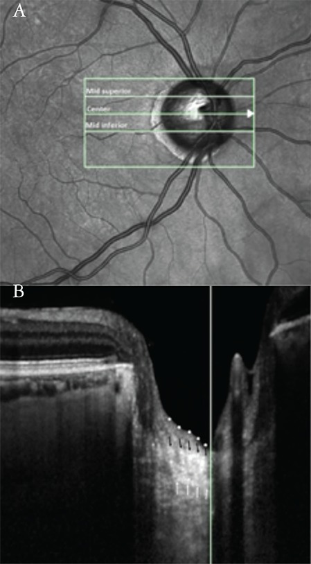
Figure 1. Simultaneous images of a pseudoexfoliation glaucoma patient. a) The measurement of prelaminar tissue thickness and laminar thickness (LT) were performed at the presumed vertical center of each of the 3 B-scans (mid-superior, center, mid-inferior). The short vertical line crossing the center horizontal line corresponds to the long white vertical line in the next image. B) The image shows a horizontal cross-sectional B-scan of the optic nerve head at the center line. The vertical white line marks the vertical center of the optic nerve head. The borders of the highly reflective region were accepted as the borders of the lamina cribrosa (LC); white arrows indicate the posterior borders and black arrows indicate the anterior borders of the LC. LT was defined as the distance between the anterior and posterior LC borders. Prelaminar tissue was defined as the reflective field on the anterior margin of the LC. White dots delineate the anterior borders of the prelaminar tissue.

