
Figure 2.
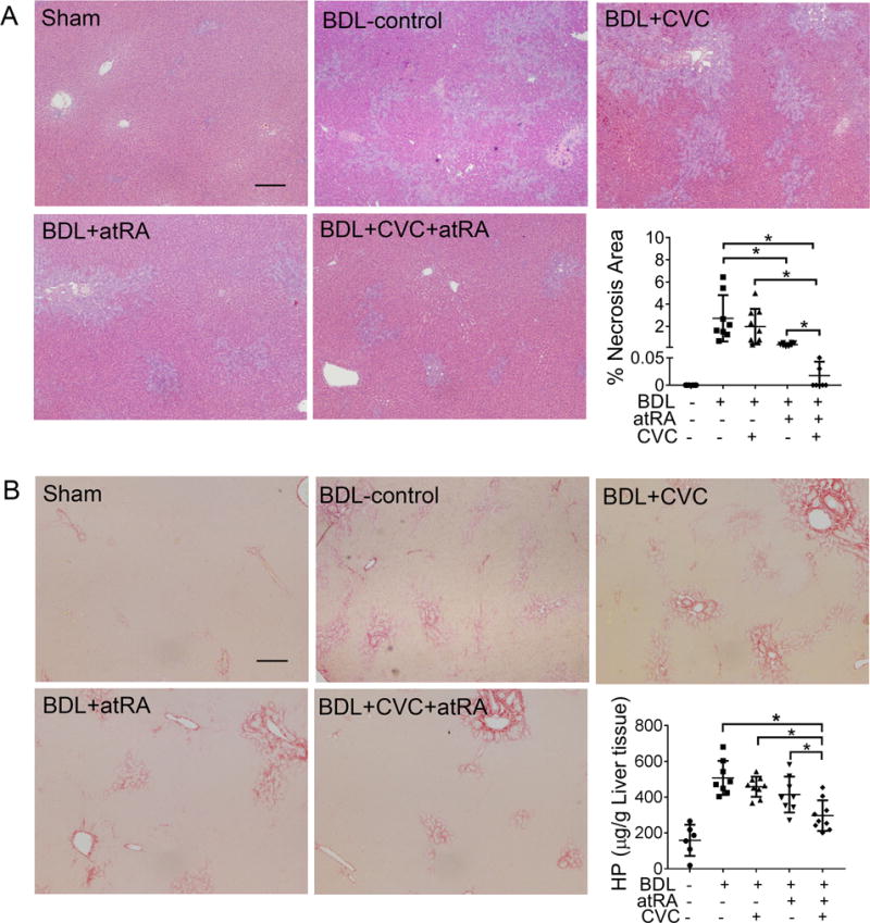
CVC in combination with atRA significantly reduced liver injury in BDL rats. (A) representative photomicrographs of hematoxylin and eosin-stained liver histology and quantitative measurement of necrotic area in all livers; (B) representative images of liver section stained with Sirius Red and detection of liver hydroxyproline content. Scale bar = 100 μm. * p<0.05, n=6-9.