Figure 7.

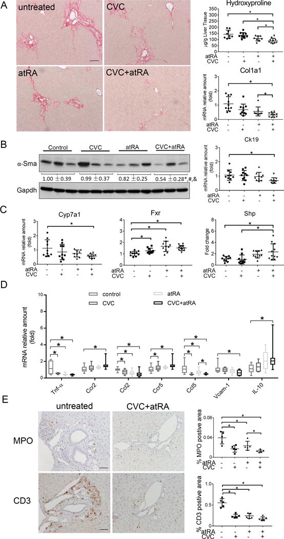
The combination treatment of atRA and CVC reduced liver injury in cholestatic Mdr2−/− mice. (A) liver Sirius Red staining, hydroxyproline content and mRNA expression of Col1a1, Scale bar = 100 μm; (B) liver protein expression of α-Sma and mRNA expression of Ck19; (C) liver mRNA expression of Cyp7a1, Fxr and Shp; (D) liver mRNA expression of inflammation related genes, i.e. Tnf-α, Ccr2, Ccl2, Ccr5, Ccl5, Vcam-1 and IL-10; and (E) immunohistochemistry for the neutrophil marker myeloperoxidase (MPO) and T cells marker CD3. Representative photomicrographs taken with a 20X objective are shown on the left, and quantitative analysis of the positive areas in the liver sections are shown on the right. Scale bar = 50 μm. * p<0.05, n=9-11.
