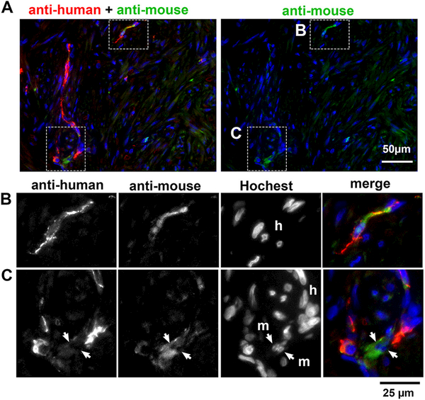
Figure 5. The origin of vascular cells in PDX.
IF assay for anti-human (red) and anti-mouse (green) PECAM1 antibodies. (A) Macro images. The areas enclosed with a dotted line are magnified in the images below. (Row B): IF assay for each antibody and Hochest staining in separate gray-scale channels and then merged demonstrates the human origin (h) of endothelial cells stained with anti-mouse PECAM1. (Row C): Likewise, a small number of mouse endothelial cells (m) are integrated in vasculature with human endothelial cells (h).

