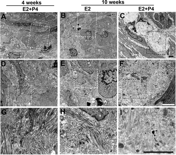
Figure 6. Analysis of collagen fibers in MED12-LMs by TEM.
TEM images of MED12-LM PDXs (MED12 c.130G>A): (A, D and G) Early growth at 4 weeks. (B, E and H) Resting at 10 weeks with E2. (C, F and I) Late growth at 10 weeks E2+P4. Images are displayed in 3 different magnifications (lower to higher from top to bottom) with bars indicating 2 μm. The area that is magnified in the row below is enclosed with a dotted line.

