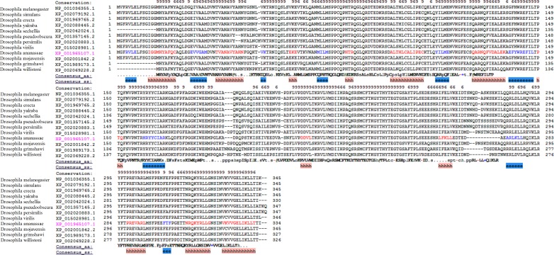
Figure 3. The multiple alignments of protein sequences retrieved from the NCBI repository with conserved domain architecture with DNMT2 methyltransferase in D. melanogaster.
The ‘e’ and ‘h’ regions represent beta strand and alpha helix consensus secondary structures from the residues in the twelve proteins.

