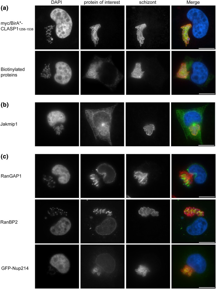
Figure 1.

Use of BioID to identify microtubule (MT)‐binding proteins and proteins of the nuclear pore complex at the schizont surface. (a) Theileria annulata‐transformed cells (TaC12) were transduced with myc‐BirA*‐CLASP11256–1538 lentivirus particles and analysed by immunofluorescence analysis. The localisation of myc‐BirA*‐CLASP11256–1538 was analysed with anti‐myc labelling (green); anti‐TaSP (red) antibodies were used to label the schizont surface (top panel). TaC12_ myc‐BirA*‐CLASP11256–1538 cells were incubated with 50‐μM biotin prior to fixation and analysis with FITC‐conjugated streptavidin (green); the parasite surface was labelled with anti‐p104 antibodies (red). DNA is labelled with DAPI (blue). (b) The MT‐interacting protein Jakmip1 associates with the parasite surface. TaC12 cells were stained with anti‐Jakmip1 (green), the parasite was labelled with anti‐p104 (red), and host and parasite nuclei were labelled with DAPI (blue). (c) Several proteins involved in nucleo‐cytoplasmic transport were identified with BioID and tested for proximity to the T. annulata schizont surface. TaC12 cells were stained with anti‐RanGAP1 (green, top panel), anti‐RanBP2 (green, middle panel), or transfected with GFP‐Nup214 (bottom panel). The parasite was labelled with anti‐p104 (red, top, and middle panels) or anti‐TaSP (red, bottom panel), and host and parasite nuclei were labelled with DAPI (blue). Scale bar = 10 μm
