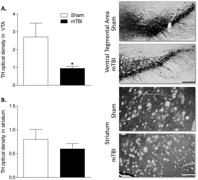
Figure 3. Juvenile mTBI reduces tyrosine hydroxylase expression in the ventral tegmental area.
A) Optical density of tyrosine hydroxylase (TH) staining revealed reduced expression in the VTA of brain injured mice, B) but no difference between mTBI and sham mice in the striatum. An asterisk (*) indicates a significant difference, p < 0.05. Representative images of the VTA and striatum are shown with scale bars of 200 μm and 100 μm respectively.

