Fig. 2.
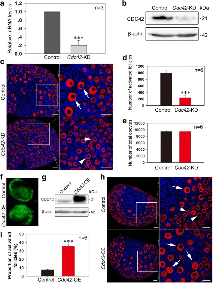
Manipulating the expression of Cdc42 controls the activation of primordial follicles. a, b Validating the efficiency of Cdc42 knockdown in cultured ovaries. Ovaries at 1 dpp were injected with Cdc42 esiRNA or scrambled siRNA and cultured in vitro for 24 or 48 h. Cdc42 mRNA and protein expression was significantly decreased in the Cdc42-KD group. c After Cdc42 esiRNA injection at 1 dpp, the transfected ovaries were cultured for six more days. Histological analysis showed a normal follicle distribution with activated follicles (arrows) in control ovaries. However, numerous primordial follicles (arrowheads) and few activated follicles were observed in the Cdc42-KD ovaries. d, e Follicle quantification in cultured ovaries showed that knockdown of Cdc42 significantly inhibited the activation of primordial follicles (P < 0.001) but had no effect on the survival of oocytes (Additional file 10: individual data values). f The ovaries at 1 dpp were transfected with empty lentivirus or a lentiviral construct expressing Cdc42 (Cdc42-OE) for 2 or 6 days in vitro. Green fluorescence from the GFP reporter was observed in ovaries following 2 days of lentiviral infection, demonstrating a satisfactory efficiency of Cdc42-OE. g Western blot showed that CDC42 expression was obviously upregulated in Cdc42-OE ovaries after 2 days of culture. h Morphological analysis showed that ovaries in the Cdc42-OE group exhibited more activated follicles (arrows) than the control. Ovaries were stained for the oocyte marker DDX4 (red). Nuclei were dyed with a Hoechst counter-stain (blue). i Quantification of ovarian follicles showed a significant increase of activated follicles (34.43 ± 2.34%) in Cdc42-OE ovaries compared to controls (8.06 ± 0.80%) (Additional file 10: individual data values). The experiments were repeated at least three times, and representative images are shown. Scale bars, 50 μm

