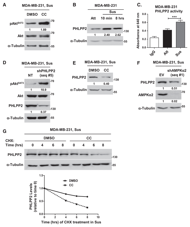
Figure 3.
AMPK promotes Akt dephosphorylation in suspension via PHLPP. A and B, Representative immunoblots of MDA-MB-231 cells treated with DMSO or compound C (CC) and subjected to suspension (Sus) for 8 hours (n = 5; A), and cells grown in adherent (Att) or suspension (Sus) condition for 10 minutes and 8 hours (n = 3; B). C, Phosphatase assay performed with immunoprecipitated PHLPP2; IgG was used as control; n = 4. Error bars, mean ± SEM. D–G, Representative immunoblots of MDA-MB-231 cells harvested under conditions detailed below. D, Cells stably expressing nontargeting shRNA (NT) or shPHLPP2 (seq #5) and subjected to suspension (Sus) for 8 hours; n = 4. E, Cells treated with DMSO or compound C (CC) were subjected to suspension (Sus) for 8 hours; n = 3. F, Adherent cells stably expressing pTRIPZ empty vector (EV) or shAMPKα2 (seq #1) were induced with doxycycline for 48 hours, followed by suspension for 8 hours; n = 3. G, Adherent cells pretreated with DMSO or AMPK inhibitor (CC) were treated with cycloheximide (CHX) for 20 minutes, followed by suspension culture (Sus) for indicated time points; n = 3. Graph represents quantification of PHLPP2. All values represent densitometric analyses of Western blots to quantify relative levels of specified proteins.

