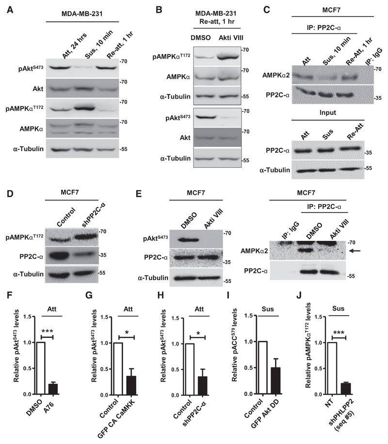
Figure 5.
Matrix reattachment leads to Akt-dependent repression of AMPK activity. A–E, Representative immunoblots of cells harvested under conditions detailed below. A, MDA-MB-231 cells cultured in attachment (Att), suspension for 10 minutes (Sus), or allowed to reattach (Re-att); n = 3. B, MDA-MB-231 cells subjected to 10 minutes of suspension (Sus) were allowed to reattach (Re-att) in the presence of DMSO or Akt inhibitor; n = 3. C, Lysates of MCF7 cells cultured in conditions of attachment (Att), suspension (Sus), and reattachment (Re-att) were immunoprecipitated (IP) with control IgG or anti–PP2C-α antibodies and analyzed by immunoblotting. The input represents 2% of the whole-cell lysate used for each immunoprecipitation; n = 3. D, Adherent MCF7 cells transfected with vector control (pLKO.1) or shPP2C-α; n = 3. E, Lysates of adherent MCF7 cells treated with DMSO or Akt inhibitor were immunoprecipitated and analyzed as described in C; n = 3. F–J, Graphs represent densitometric quantification of immunoblots (Supplementary Figs. S7A-S7E) for relative phospho-protein levels from cells harvested under conditions detailed below: F, Adherent MDA-MB-231 cells treated with DMSO or AMPK activator (A76; also see Supplementary Fig. S7A); n = 3. G, Adherent MDA-MB-231 cells transfected with control vector pEGFP or GFP-tagged constitutively active CaMKK (GFP CA CaMKK; also see Supplementary Fig. S7B); n = 3. H, Adherent MCF7 cells transfected with vector control (pLKO.1) or shPP2C-α (also see Supplementary Fig. S7C); n = 2. I, MCF7 cells transfected with GFP or GFP-HA-Akt-T308D S473D and subjected to suspension (8 hours; also see Supplementary Fig. S7D); n = 2. J, MDA-MB-231 cells stably expressing nontargeting shRNA (NT) or shPHLPP2 (seq #5) and subjected to suspension (48 hours; also see Supplementary Fig. S7E); n = 3. Error bars, mean ± SEM.

