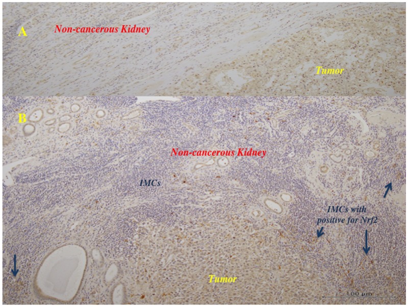
Figure 5. Immunohistochemistry in the primary tumor tissues for Nrf2.
The tumor cells showed positive staining for anti-Nrf2 antibody. (A) (x100): In the tumors with lower histological grade (Fuhrman grade 1/2), many of the tumor cells showed weak to moderate reaction for anti-Nrf2 antibody, and the surrounding non-tumor tissues showed negative to very weak reaction. There were little infiltrating immune (mononuclear) cells (IMCs). (B) (x100): In the tumors with higher histological grade (Fuhrman grade 3), much of the tumor cells showed moderate to strong brown staining, and some IMCs showed strong reaction (arrows).

